
| PRODUCTS | |
Our assortment of fine tools for molecular biology is constantly growing.
So far we offer ten products for various uses in molecular biology for a price you wouldn't believe. Please refer to one of the links below:

|
NOWA
Chemiluminescent Western This new detection kit for HRP-conjugated antibodies offers a sensitivity far beyond anything known for a price you haven't even dreamed of! |

|
Enlight
Fluorographic Enhancer This non-toxic aqueous solution allows the enhancement of low-energy isotopes within acrylamide gels. Exposure times can thus be reduced significantly. Works better than other known products on the market! |

|
Silver Surfer
Single Solution Silver Stain This single-solution staining kit combines the sensitivity of silver stain with speed end ease of use. Believe it or not - staining takes place in one single solution and is completed just 30 minutes after fixing. |

|
Snow White
Reversible Zinc Stain This widely unknown staining protocol beats everything in terms of speed. In just 10 minutes after detaching your gel plates you can document your gel! Sensitivity is close to silver and the staining is reversible! Apply your destained gel to whatever procedure you want. |

|
Deep Blue
Colloidal Coomassie Coomassie staining is still the most common visualisation for protein SDS gels. This kit allows a very sensitive coomassie stain that requires no subsequent destaining! Get you results quicker (1 hour) and keep the odor and atmosphere in your lab clean. |

|
Bradford
Protein Quantification It's the classic colorimetric assay for measuring total protein concentration. Apart from a price that only we do offer, Bradford comes with a protocol that offers an increased linearity of OD to concentration ratio. |

|
NuClean
Silica Matrix DNA Purification Like other kits for purifying nucleic acids from agarose gels or solutions NuClean is based on the use of a powdered silica matrix. The price per reaction however is incomparable with other kits on the market! Comes with 4 ml of silica matrix suspension. |

|
pEG-His1
His-Tagged Cloning Vector We now offer our first vector. Due to its exceptional tightness this expression vector is especially suitable for the expression of toxic genes in E.coli. An optional C-terminal His-tag is included for easy purification of full-length only. |

|
GLOW
Microscopy Mounting Medium This mounting medium for immunofluorescent microscopy substantially decreases bleaching of fluorochromes under UV radiation. Since it also solidifies on the slide the use of nail polish for slide fixation is history. |

|
T.Wax
HotStart PCR Beads Ever tried hot-start PCR to increase your specificity? T.Wax are easy-to-use wax beads that allow a contamination-free and reproductive application of this powerful method. |
 NOWA
Chemiluminescent Western
NOWA
Chemiluminescent Western
NOWA is a highly sensitive detection system for
HRP-conjugated antibodies in the picogramm range. The kit is
based on the emission of light during a reaction which is started
be free oxygen produced by the enzymatic activity of HRP.
Using a newly developed enhancement cascade the light emission
is amplified by a factor of at least one thousand. The NOWA kit
contains two solutions that are combined and used to incubate the
nitrocellulose or PVDF filter in. After one minute of incubation
time the filter is ready to be exposed to an x-ray film.
Typical exposure times range between 10 seconds and
5 minutes. The light emitting reaction continues for
about 1 hour to allow as many exposures as you may need.
The kit contains 2 x 250 ml and is sufficient
for 10 000 cm2 (1 m2!)
of NC or PVDF membrane resulting in a price of less than
0.50 EURO per mini gel blot!
| Article | Quantity | Catalog No. | Price |
| NOWA | 2 x 250 ml | W001 0250 | EURO 79.00 |
Literature:
Schneppenheim, R. et al. 1991. Luminography - a highly sensitive visualization method for electrophoresis. Electrophoresis. 12(5): 367-372.
  Light emission of NOWA
  versus popular Brand X
  during two hours of
  reaction time.
  (RLU=relative light units)
 Enlight
Fluorographic Enhancer
Enlight
Fluorographic Enhancer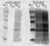
 Silver Surfer
Single Solution Silver Stain
Silver Surfer
Single Solution Silver Stain
 Snow White
Reversible Zinc Stain
Snow White
Reversible Zinc Stain
 Deep Blue
Colloidal Coomassie
Deep Blue
Colloidal Coomassie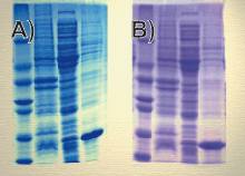
 Bradford
Protein Quantification
Bradford
Protein Quantification
 NuClean
Silica Matrix DNA Purification
NuClean
Silica Matrix DNA Purification
 pEG-His1
His-Tagged Cloning Vector
pEG-His1
His-Tagged Cloning Vector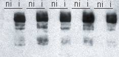
 GLOW
Microscopy Mounting Medium
GLOW
Microscopy Mounting Medium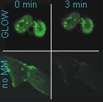
 T.Wax
HotStart PCR Beads
T.Wax
HotStart PCR Beads